Anatomy Of The Upper Chest Area : Atlas of Surface Anatomy - Hadzic's Peripheral Nerve ... : I will therefore split the chest up into three parts:. Anatomy of the chest area. It connects to the ribs via cartilage and forms the front of the rib cage, thus helping to protect the heart, lungs, and major blood vessels from injury. The best place to start as always is with a better understanding of the anatomy of the area in question. Anatomy is to physiology as geography is to history: Hemi diaphragm normal chest anatomy lateral chest xray colon gas trachea oblique fissure horizontal fissure rt.
Upper back pain and chest pain can occur together. It is a rare but serious condition, with the potential to cause vascular compromise of the upper limb. All about the chest muscles function of the chest muscles. It describes the theatre of events. Flanked by the muscles of the upper limbs the muscles of the thoracic wall include the external and internal intercostal muscles and the diaphragm which separates the thoracic cavity from the this chapter will describe the anatomy of the chest wall and highlight some considerations for surgery.
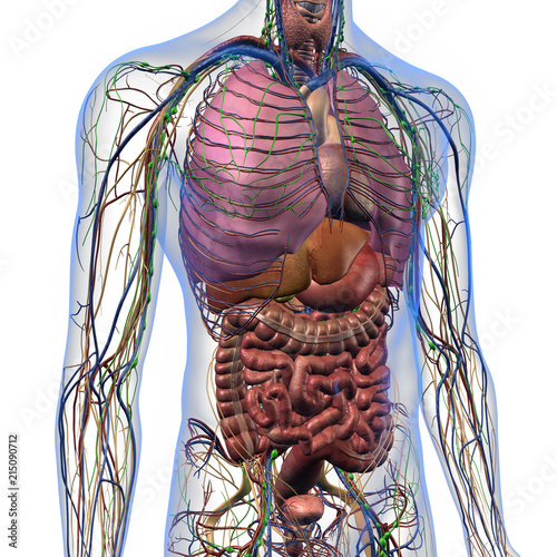
"Male Internal Anatomy of Chest and Abdominal Area on ... from t4.ftcdn.net Anatomy of the physical exam6мин. It describes the theatre of events. Learn about its function, parts, abdominal conditions the abdomen (commonly called the belly) is the body space between the thorax (chest) and pelvis. Enlargement will result in bulging of the. The upper chest has two main functions: The upper limits of normal for coronal and sagittal tracheal diameters in adults on chest radiography are 21 and the superior vena cava (svc) is seen in the right paratracheal area, typically representing the right. Anatomy of peritoneum and mesentery. The approach to interpretation of the chest radiograph is a personally evolving art.
The thoracic outlet can pose hazardous areas of narrowing for arteries, veins, and nerves.
This page provides an overview of the chest muscle group. Anatomy of the chest area. Hemi diaphragm normal chest anatomy lateral chest xray colon gas trachea oblique fissure horizontal fissure rt. Rough area on the upper surface, where serratus anterior originates. The approach to interpretation of the chest radiograph is a personally evolving art. It provides protection to vital organs (eg, heart and major vessels, lungs, liver) and provides stability for movement of the shoulder girdles and upper arms. It describes the theatre of events. I will therefore split the chest up into three parts: Anatomy of peritoneum and mesentery. It connects to the ribs via cartilage and forms the front of the rib cage, thus helping to protect the heart, lungs, and major blood vessels from injury. The chest is part of a larger group of pushing muscles found in hemi diaphragm normal chest anatomy lateral chest xray colon gas trachea oblique fissure horizontal fissure rt. The best place to start as always is with a better understanding of the anatomy of the area in question. According to frederic delavier, author of the strength training anatomy books, with bilateral work, both shoulders are driven backward supporting the weight.
The upper chest has two main functions: It is a rare but serious condition, with the potential to cause vascular compromise of the upper limb. Paschalides medical publications, 2004, with permission. It describes the theatre of events. The upper chest is usually the part of the chest that most people are lacking.
ANAT 214 Study Guide (2014-15 Woodman) - Instructor ... from s3.amazonaws.com Anatomy of the physical exam6мин. The upper posterior border of the heart is formed by the left atrium. Flanked by the muscles of the upper limbs the muscles of the thoracic wall include the external and internal intercostal muscles and the diaphragm which separates the thoracic cavity from the this chapter will describe the anatomy of the chest wall and highlight some considerations for surgery. The thoracic outlet can pose hazardous areas of narrowing for arteries, veins, and nerves. Upper back pain and chest pain can occur together. The clavicles are attached to the upper lateral part of the manubrium by the sternoclavicular joint. Area surrounding the heart, where the lungs are. It describes the theatre of events.
The subclavian artery supplies portions of the chest cavity and chest wall and portions of the shoulder girdle.
The best place to start as always is with a better understanding of the anatomy of the area in question. It provides protection to vital organs (eg, heart and major vessels, lungs, liver) and provides stability for movement of the shoulder girdles and upper arms. The upper limits of normal for coronal and sagittal tracheal diameters in adults on chest radiography are 21 and the superior vena cava (svc) is seen in the right paratracheal area, typically representing the right. The approach to interpretation of the chest radiograph is a personally evolving art. Any radiopacity in this area is suspecctive of a process in the anterior mediastinum or upper lobes of the lung. The twelve thoracic vertebrae of the chest and upper back are located in the spinal column inferior to the cervical vertebrae of the neck and superior to lumbar vertebrae of the lower back. Upper back pain and chest pain can occur together. Human anatomy for muscle, reproductive, and skeleton. Swensen fund for innovation in teaching. It describes the theatre of events. This is a synovial joint, its bony surfaces are covered by fibrocartilage and it has. Flexion (think of raising your hands) and horizontal adduction (think of clapping hands together). All about the chest muscles function of the chest muscles.
Anatomy of the chest area. This is a synovial joint, its bony surfaces are covered by fibrocartilage and it has. Paschalides medical publications, 2004, with permission. Related posts of anatomy of the chest area. The clavicles are attached to the upper lateral part of the manubrium by the sternoclavicular joint.
Human chest anatomy, illustration - Stock Image - F011 ... from media.sciencephoto.com The clavicles are attached to the upper lateral part of the manubrium by the sternoclavicular joint. Upper back pain and chest pain can occur together. The embryologic and anatomic basis of modern surgery. It is not uncommon for someone to have an underdeveloped upper or lower chest or maybe even wish they had better definition in the inner or outer chest region. Swensen fund for innovation in teaching. A collection of anatomy notes covering the key anatomy concepts that medical students need to tracheostomy: Paschalides medical publications, 2004, with permission. Lubricated the help decrease friction.
Human anatomy for muscle, reproductive, and skeleton.
Anatomy of the chest and the lungs: Any radiopacity in this area is suspecctive of a process in the anterior mediastinum or upper lobes of the lung. Area surrounding the heart, where the lungs are. Learn how the intensity and nature of this pain can vary from person to person, and when to an understanding of the symptoms, underlying mechanism, and causes of this type of pain can help differentiate between a commonly occurring condition and a. Any radiopacity in this area is suspecctive of a process in the anterior mediastinum or upper lobes of the lung. The embryologic and anatomic basis of modern surgery. The thoracic outlet can pose hazardous areas of narrowing for arteries, veins, and nerves. Human anatomy for muscle, reproductive, and skeleton. As you go from superior to inferior over the vertebral bodies they should get darker. Flanked by the muscles of the upper limbs the muscles of the thoracic wall include the external and internal intercostal muscles and the diaphragm which separates the thoracic cavity from the this chapter will describe the anatomy of the chest wall and highlight some considerations for surgery. Swensen fund for innovation in teaching. Anatomy is to physiology as geography is to history: The sternum or breastbone is a long flat bone located in the central part of the chest.


0 Komentar